Anatomy Of The Shoulder: Understanding The Structures Involved
The shoulder is a marvel of anatomical complexity, offering remarkable flexibility and range of motion. However, this versatility also makes it vulnerable to injury and pain. Understanding the intricate structures that compose the shoulder joint is crucial for grasping its function, recognizing potential issues, and pursuing effective treatment. In this article, we delve into the anatomy of the shoulder, exploring its key components and their roles in facilitating movement and stability.
To Know More About It please Click Here
An Overview of Shoulder Anatomy
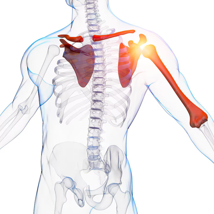
The shoulder is a ball-and-socket joint formed by the articulation of three bones: the humerus (upper arm bone), the scapula (shoulder blade), and the clavicle (collarbone). Unlike the hip joint, which is characterized by deep bony sockets, the shoulder joint sacrifices stability for mobility.
Key Structures
- Glenohumeral Joint:
- This is the primary joint of the shoulder, where the rounded head of the humerus articulates with the shallow socket of the scapula, known as the glenoid cavity.
- The joint capsule, a sleeve-like structure, surrounds the glenohumeral joint, providing stability and containing synovial fluid for lubrication.
- Ligaments, including the glenohumeral ligaments and the coracohumeral ligament, reinforce the joint capsule, helping to prevent excessive movement.
- Rotator Cuff:
- Comprised of four muscles and their tendons—the supraspinatus, infraspinatus, teres minor, and subscapularis—the rotator cuff plays a vital role in stabilizing the shoulder and facilitating overhead movements.
- These muscles originate from the scapula and attach to the humerus, forming a cuff that encircles the glenohumeral joint.
- The rotator cuff tendons pass beneath the acromion, a bony projection of the scapula, through a space called the subacromial space. This area is prone to impingement, a common cause of shoulder pain.
- Acromioclavicular (AC) Joint:
- Located at the junction of the acromion (part of the scapula) and the clavicle, the AC joint allows for movements such as raising the arm overhead.
- Ligaments, including the acromioclavicular ligament and the coracoclavicular ligaments, stabilize the AC joint.
- Injuries to the AC joint, such as sprains or separations, can result from trauma or overuse.
- Scapulothoracic Joint:
- The scapula rests on the posterior aspect of the rib cage and moves along the thoracic wall as the arm moves.
- Muscles attached to the scapula, including the trapezius, serratus anterior, and rhomboids, play a crucial role in stabilizing and positioning the scapula during shoulder movements.
Understanding Function and Dysfunction
The intricate interplay of these structures allows for the remarkable range of motion exhibited by the shoulder. However, this complexity also renders the shoulder vulnerable to various conditions, including rotator cuff tears, shoulder impingement, shoulder dislocation, and osteoarthritis.
Conclusion
The shoulder’s anatomy is a testament to the marvels of human biomechanics, enabling a wide range of movements essential for daily activities and athletic endeavors. By gaining a deeper understanding of its structures and functions, individuals can better appreciate the importance of shoulder health and take proactive measures to prevent injuries and address any issues that arise. Whether through proper posture, targeted exercises, or seeking medical guidance when needed, nurturing the well-being of the shoulder is key to maintaining a healthy and active lifestyle.
Also, Follow us on Instagram